A portable two-channel PPG
cardiovascular sensor device
Janis Spigulis*, Renars
Erts and Maris Ozols
ABSTRACT
A
portable sensor device for simultaneous detection
and processing of skin-remitted optical signals from
any two sites of the body has been developed and tested. The photoplethysmography (PPG) principle was applied to follow
the dilatation and contraction of skin blood vessels during the cardiac cycle.
The newly developed two-channel
approach allows to estimate the vascular blood flow
resistance by analysis of time shifts between the PPG pulses detected at
different body sites. Potential of the sensor device for express-assessment of human cardio-vascular condition
and for body fitness tests has been demonstrated.
Keywords: Photoplethysmography, optical bio-sensing, cardio-vascular
assessment. fitness control.
1.
INTRODUCTION
The human’s cardio-vascular
condition can be assessed both invasively and non-invasively. Photoplethysmography (PPG) is a non-invasive method for
studies of the blood volume pulsations by detection and analysis of the tissue
back-scattered or absorbed optical radiation. Blood pumping and transport
dynamics can be monitored at different body locations - fingertip, earlobe,
forehead, forearm, etc. – with relatively simple and convenient PPG contact
probes. PPG technique potentially may become a routine everyday tool for
express diagnostics and early screening of cardio-vascular pathologies, as well
as for self-monitoring of the vascular condition. Tele-diagnostics by means of PC-connections
via Internet or LAN is another area where advanced PPG-technology becomes very
important.
In general, each recorded PPG
pulse contains information, useful for cardio-vascular assessment. However, all
detected heartbeat pulses are not equal – the PPG signal amplitude, baseline
and period are changing with time 1. The real bio-signals are
fluctuating around their mean value that we call single-period photoplethysmography (SP-PPG) signal. It can be
determined by averaging a sequence of about 50…80 PPG pulses by means of
specific algorithms and PC-processing programmes 2 - 4.
Our first wheel-table SP-PPG
fingertip sensor devices 2, 3 had undergone several series of
clinical tests in laboratory, classroom and hospital environments. Analysis of
signals taken from numerous volunteers had lead to conclusion that each person
has his/her specific shape of the mean SP-PPG signal,
and this "PPG-fingerprint" reflects the individual's cardio-vascular
condition. This observation confirmed the diagnostic potential of the developed
devices; however, their hospital bed-site and field applications were limited
due to considerable weight/size of the table-top computer placed on the
wheel-table and dependency on the wall power plugs, therefore a smaller sensor
model based on a lap-top computer was created as the next step 4.
Recently
a portable version of two-channel PPG sensor device was developed and tested.
The sensor comprises a set of universal optical contact probes, electronic
converters and a laptop computer with specially designed software providing
real-time display, processing and storage of the PPG signals recorded at both
channels. The
newly developed two-channel approach is more informative – on-line monitoring
of two body sites not only doubles the data flow but also opens possibility to
estimate the vascular blood flow resistance. It is proportional to the
heartbeat wave propagation time - the measurable time shift between the two
corresponding PPG pulses detected at two different body sites. Our studies confirmed
the promising potential of this methodology and sensor design for easy and fast cardio-vascular assessment
and for body fitness tests.
*) E-mail: janispi@latnet.lv,
tel/fax: +371 7228249
2. DESIGN OF THE TWO-CHANNEL PPG SENSOR
DEVICE
The PPG sensor device consists of
a set of optical contact probes (two of them are used simultaneously during the
measurements), the bio-signal acquisition circuit, and the lap-top computer
with specially developed software. All equipment is placed in a hand-held case 4
of size 44x32x9 cm and weight 4.1
kg; it is battery-powered and can operate up to 3 hours without recharging.
Each optoelectronic contact probe
emits cw-radiation into the skin tissues and detects
the back-scattered radiation; its separated AC-component precisely reflects the
skin blood volume pulsations. Each contact probe comprises a GaAs emitting diode (diameter of the emitting area ~2 mm,
radiant power ~10 mW, peak wavelength ~940 nm, the estimated
mean penetration depth under the skin surface ~2 mm), and a Si
photodiode with square detection area ~5x5 mm. Both diodes are closely mounted
on a soft plastic pillow and fixed onto the measurement site by means of a
sticky band – see Fig. 2,a. The band length is adjusted for the fingertip
measurements (Fig. 2, b); also a standard pulse oximetry
finger-clip was adapted for our measurements, maintaining the same emitter-detector
geometry as in the band-mounted probe.
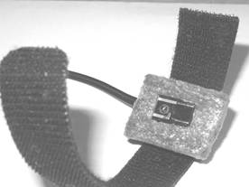
a b
Fig. 1. The PPG
contact probe (a) and its application for monitoring of blood pulsations in
fingertip (b).
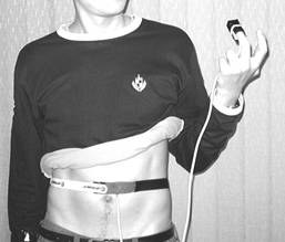
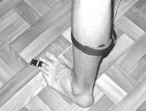
a b
Fig. 2. Two-channel PPG
measurements: placing the contact probes at fingertip and belly (a), and at
calf and toe (b).
Advantage of the band-based probe
design is the possibility of its flexible extension by means of spare sticky
bands, so allowing to take the PPG measurements from various locations of the
body, e.g. fingertip, belly, calf, toe (Fig. 2).
Special software was developed for the PPG
bio-signal acquisition, processing and data storage related to both input
channels, offering the following options:
·
Filling the first window for patient data - name,
age, gender, complains, doctor’s comments, etc.;
·
Pre-setting the measurement time schedule;
·
The PPG signal real-time display on the monitor;
·
Signal clean-up (special filtering algorithm) and
calculation of the mean single-period PPG (SP-PPG) signal shape;
·
Calculation of specific cardio-vascular parameters
for the registered signals - heartbeat rate, anacrota rise-time, time delay
and relative amplitude of the secondary peak (dycrotic notch), time-delay
between two corresponding PPG signals at both channels, etc.;
·
Display of the corresponding PPG parameter set with
subsequent cardio-vascular assessment results;
·
Storage of the
measurement/assessment data.
The data sampling rate 100 s -1 was
usually chosen; it can be increased up to 950 s -1 for special cases
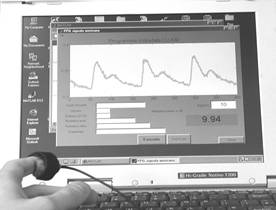
when higher time resolution is needed. The device monitor screenshot during two-channel
PPG measurements is presented at Fig. 3.
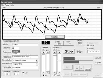
Fig. 3. The monitor screenshot during
two-channel PPG measurements.
3. APPLICATION 1: CARDIO-VASCULAR TELE-MONITORING
VIA INTERNET
In
order to check suitability of the new PPG sensor device for telemedicine needs,
an Internet transmission experiment was carried out, using the WinSock
interface between the TCP/IP protocol
and our Windows measurement program. The measured PPG signal values of both
channels were first packed together with the corresponding time reading value,
and then the packages were transmitted in real time with rate ~ 6 Kb/s via the Realtek 56 K modem. The signals were further
registered by another computer (with 225 MHz processor and appropriate
software) that was connected to Internet via the radio-link. Practically unchanged signal data were received in
few seconds (Fig. 4). Full analysis of the transmitted and received data gave
the signal loss probability less than 0.7 % in this case.
Consequently,
the developed sensor device and software can be used in telemedicine systems,
e. g. for patient home monitoring under doctor’s tele-control.

Fig.
4. The telemedicine experiment: comparison of the two-channel PPG signals
transmitted via Internet.
4. APPLICATION 2: CARDIO-VASCULAR MONITORING
DURING THE FITNESS TESTS
Advantage of the newly developed
two-channel approach is availability of additional diagnostic information on
the vascular blood flow resistance – it can be obtained by measuring the
heartbeat wave propagation time between two body contact sites as the time
shift between the two corresponding PPG pulses. For example, Fig. 5 illustrates
distinct time delay between the PPG signals recorded simultaneously at the left
fingertip and left toe (a), and at the left toe and belly (b). Time resolution
of the device is high enough to provide reliable blood pulse wave propagation
velocity estimations for diagnostic needs.
b a
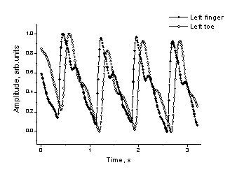

Fig. 5. Time-shifted PPG signals detected at different body sites:
a – finger and toe, b – toe and belly.
We performed series of PPG measurements before and
after intensive physical exercises aiming to follow the cardio-vascular
relaxation process that reflects adaptability of the body to physical loads.
The monitored volunteers – about 200 in total - were persons of different ages,
genders and training background. We studied the PPG signals detected at
fingertip only and at fingertip simultaneously with signals detected over the
carotid artery on the neck. The goal of this study was to find out specific
exercise-induced features of bio-signals, giving evidence on possible
cardio-vascular disorders of the monitored person.
In single-channel experiments, such evidence might
be sharp spasmatic peaks in the fingertip PPG signals
appearing for some persons immediately after intensive running (Fig. 6, a -
upper curve) or after a short relaxation time (Fig. 6, b - the next curve from
the top). Probably they can also serve as markers of the cardio-vascular
adaptation to physical loads.


a b
Fig. 6. The spasmatic
peaks observed in fingertip PPG signals after intensive running.
The two-channel PPG methodology can ensure more
informative fitness tests – in addition to the traditional physiological
parameters (e. g. pulse rate, blood pressure), monitoring of vascular
resistance changes reflected as the changes in time shifts of corresponding PPG
signal pulses is also available. We
performed a number of measurements during a complex fitness tests comprising 1
minute horizontal relax, 1 minute standing position, 3 minutes metronome-controlled
stepping up and down (the Harvard step-test 5), and 5 minutes relax in
sitting position. The dual-channel PPG signals were recorded continuously all
10 minutes at two body locations – the Carotid artery on the neck and the left
middle fingertip. As result, several functional parameters were calculated 6
- the heartbeat pulse rate changes (from time intervals between the PPG peaks
in each channel), the dominant pulse rate oscillation frequencies (by Fourier
analysis), the recreation time after the exercise (by calculating the time
constant of exponential pulse rate decay), as well as the pulse wave propagation time between
the two contact sites (from time shifts between the corresponding PPG pulses
detected in both channels).
As example, the pulse rate changes and the 2-channel
PPG time shifts for the same person (male, 23 years) are presented on Fig. 7;
the mean SP-PPG fingertip signal shapes for this person are presented on Fig.
8. Following all the test phases, there are notable changes not only in pulse
rate, but also in time-shifts between the PPG pulses in both channels, so
indicating that the vascular resistance was changing. Such sensitivity of time shifts we observed
only for few persons; most of the monitored volunteers had roughly the same
time shifts at all phases of this test.
One more advantage of the two-channel approach is
possibility to verify the suspicious features of signals at one channel by
comparing with those detected at the other channel. For instance, Fig. 9 shows
the after-exercise heart arrhythmia case convincingly detected simultaneously
at both channels.
a

2. 1.
b

Fig 7. Recorded pulse
rate changes during the fitness step-test (a), and the corresponding changes in
time
delay
between the PPG signals detected at two body sites - Carotid artery and left
fingertip (b).

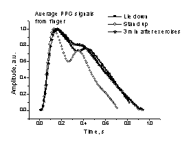
Fig. 8.
The averaged single-period PPG signals Fig. 9. Heart
arrhythmia, detected at both channels.
at different
phases of the fitness test.
5. SUMMARY
·
A
small-size portable PPG sensor device (44x32x9 cm,
4.1 kg, battery-powered) with two simultaneous detection channels is
constructed and tested.
·
The
newly developed sensor is adapted for use in tele-medicine,
e. g. for distant cardio-vascular monitoring via Internet.
·
The
proposed two-channel PPG methodology is well suited for cardio-vascular
monitoring at steady state and during complex fitness tests; the second channel
can serve for reference and for obtaining additional physiological data.
·
In
particular, time-shift between the PPG pulses that are detected at two
different body sites provides additional information on the vascular blood flow
resistance and its changes.
·
The
portable design and presented results confirm good potential of the device for
fast non-invasive cardio-vascular monitoring and mass screening at different
conditions, including bed-site and field environments.
ACKNOWLEDGMENTS
The
authors would express their sincerest thanks to Prof. Juris
Aivars and Dr. Indulis Kukulis for their valuable clinical comments. Financial
support from Latvian Council of Science (grant # 01.0067) and Latvian Ministry
of Education and Science (grant # TOP 02-13) is highly appreciated.
REFERENCES
1.
M.
Nitzan, H. de Boer, S. Turivnenko
et al., “Power spectrum analysis of spontaneous fluctuations in the photoplethysmographic signal”, J. Bas. Clin. Physiol.
Pharmacol.,
5, No. 3-4, pp. 269-276, 1994.
2.
J.
Spigulis, G. Venckus, M. Ozols, “Optical sensing for early cardiovascular
diagnostics”, Proc. SPIE 3911, pp. 27-31, 2000.
3.
J.
Spigulis, I. Kukulis, E. Fridenberga, G. Venckus,
“Potential of advanced photoplethysmography sensing
for non-invasive vascular diagnostics and early screening”, Proc. SPIE 4625, pp. 38-43, 2002.
4.
J.
Spigulis, M. Ozols, R. Erts, K. Prieditis, “A portable
device for optical assessment of the cardiovascular condition”, Proc. SPIE
5123, pp. 313-319, 2003.
5.
L. Brouha, A. Graybiel, C. W. Heath, “The
step test. A simple method of measuring physical fitness for hard muscular work
in adult men”, Rev. Canadian Biol., 2, pp. 86-92, 1943.
6.
J.
Spigulis, R. Erts, V. Bernhards, “Optics for fitness assessment: potential of
two-channel photoplethysmography techniques”, Abstr. of “Northern
Optics 2003”,